Placeholder Content Image

What the “father of the cell phone” wants you to know

<p dir="ltr">The inventor of the mobile phone has shared his candid opinion about the obsession with smart devices. </p> <p dir="ltr">Martin Cooper, an American engineer dubbed the “father of the cell phone”, invented the very first mobile phone 50 years ago in 1973. </p> <p dir="ltr">Back then, the weighty block of wires and circuits were only used to make calls, a far cry from having the world at your fingertips with smartphones today. </p> <p dir="ltr">Cooper believes that despite all the good that can come from modern technology, the world has become a little obsessed with smart devices. </p> <p dir="ltr">“I am devastated when I see somebody crossing the street and looking at their cell phone. They are out of their minds,” the 94-year-old told AFP from his office in Del Mar, California.</p> <p dir="ltr">“But after a few people get run over by cars, they’ll figure it out,” he joked.</p> <p dir="ltr">Mr Cooper also indulges in the latest gadgets, as he wears an Apple Watch and uses a top-end iPhone, flicking intuitively between his email, photos, YouTube and the controls for his hearing aid.</p> <p dir="ltr">Despite keeping up with all the latest apps, updates and upgrades, he confessed that sometimes it can all seem a little overwhelming. </p> <p dir="ltr">“I will never, ever understand how to use the cell phone the way my grandchildren and great grandchildren do,” he said.</p> <p dir="ltr">“Each generation is going to be smarter … they will learn how to use the cell phone more effectively,” he said.</p> <p dir="ltr">“Humans sooner or later figure it out.”</p> <p dir="ltr"><em>Image credits: Getty Images</em></p>

Technology

Placeholder Content Image

Long COVID: How lost connections between nerve cells in the brain may explain cognitive symptoms

<p>For a portion of people who get COVID, symptoms continue for <a href="https://www.ons.gov.uk/peoplepopulationandcommunity/healthandsocialcare/conditionsanddiseases/bulletins/prevalenceofongoingsymptomsfollowingcoronaviruscovid19infectionintheuk/6october2022" target="_blank" rel="noopener">months or even years</a> after the initial infection. This is commonly referred to as “long COVID”.</p> <p>Some people with long COVID complain of “<a href="https://theconversation.com/what-is-and-what-isnt-brain-fog-190537" target="_blank" rel="noopener">brain fog</a>”, which includes a wide variety of cognitive symptoms affecting memory, concentration, sleep and speech. There’s also growing concern about findings that people who have had COVID are at <a href="https://www.thelancet.com/journals/lanpsy/article/PIIS2215-0366(22)00260-7/fulltext" target="_blank" rel="noopener">increased risk</a> of developing brain disorders, such as dementia.</p> <p>Scientists are working to understand how exactly a COVID infection affects the human brain. But this is difficult to study, because we can’t experiment on living people’s brains. One way around this is to create <a href="https://www.nature.com/articles/s41578-021-00279-y" target="_blank" rel="noopener">organoids</a>, which are miniature organs grown from stem cells.</p> <p>In a <a href="https://www.nature.com/articles/s41380-022-01786-2.pdf" target="_blank" rel="noopener">recent study</a>, we created brain organoids a little bigger than a pinhead and infected them with SARS-CoV-2, the virus that causes COVID-19.</p> <p>In these organoids, we found that an excessive number of synapses (the connections between brain cells) were eliminated – more than you would expect to see in a normal brain.</p> <p>Synapses are important because they allow neurons to communicate with each other. Still, the elimination of a certain amount of inactive synapses is part of normal brain function. The brain essentially gets rid of old connections when they’re no longer needed, and makes way for new connections, allowing for more efficient functioning.</p> <p>One of the crucial functions of the brain’s immune cells, or <a href="https://www.ncbi.nlm.nih.gov/pmc/articles/PMC5768411/" target="_blank" rel="noopener">microglia</a>, is to prune these inactive synapses.</p> <p>The exaggerated elimination of synapses we saw in the COVID-infected models could explain why some people have cognitive symptoms as part of long COVID.</p> <p><strong>Parallels with neurodegenerative disorders</strong></p> <p>Interestingly, this pruning process is believed to go awry in several disorders affecting the brain. In particular, excessive elimination of synapses has recently been linked to <a href="https://www.nature.com/articles/s41593-018-0334-7" target="_blank" rel="noopener">neurodevelopmental disorders</a> such as <a href="https://www.nature.com/articles/s41593-018-0334-7" target="_blank" rel="noopener">schizophrenia</a>, as well as <a href="https://www.frontiersin.org/articles/10.3389/fncel.2019.00063/full" target="_blank" rel="noopener">neurodegenerative disorders</a> such as Alzheimer’s and Parkinson’s disease.</p> <p>By sequencing the RNA of single cells, we could study how different cell types in the organoid responded to the virus. We found that the pattern of genes turned on and off by the microglia in our COVID-infected organoids mimicked changes seen in neurodegenerative disorders.</p> <p>This may go some way in explaining the link between COVID and the risk of developing certain neurological disorders.</p> <figure class="align-center "><img src="https://images.theconversation.com/files/491380/original/file-20221024-17-9wi5pg.png?ixlib=rb-1.1.0&amp;q=45&amp;auto=format&amp;w=754&amp;fit=clip" sizes="(min-width: 1466px) 754px, (max-width: 599px) 100vw, (min-width: 600px) 600px, 237px" srcset="https://images.theconversation.com/files/491380/original/file-20221024-17-9wi5pg.png?ixlib=rb-1.1.0&amp;q=45&amp;auto=format&amp;w=600&amp;h=338&amp;fit=crop&amp;dpr=1 600w, https://images.theconversation.com/files/491380/original/file-20221024-17-9wi5pg.png?ixlib=rb-1.1.0&amp;q=30&amp;auto=format&amp;w=600&amp;h=338&amp;fit=crop&amp;dpr=2 1200w, https://images.theconversation.com/files/491380/original/file-20221024-17-9wi5pg.png?ixlib=rb-1.1.0&amp;q=15&amp;auto=format&amp;w=600&amp;h=338&amp;fit=crop&amp;dpr=3 1800w, https://images.theconversation.com/files/491380/original/file-20221024-17-9wi5pg.png?ixlib=rb-1.1.0&amp;q=45&amp;auto=format&amp;w=754&amp;h=425&amp;fit=crop&amp;dpr=1 754w, https://images.theconversation.com/files/491380/original/file-20221024-17-9wi5pg.png?ixlib=rb-1.1.0&amp;q=30&amp;auto=format&amp;w=754&amp;h=425&amp;fit=crop&amp;dpr=2 1508w, https://images.theconversation.com/files/491380/original/file-20221024-17-9wi5pg.png?ixlib=rb-1.1.0&amp;q=15&amp;auto=format&amp;w=754&amp;h=425&amp;fit=crop&amp;dpr=3 2262w" alt="" /><figcaption><span class="caption">A brain organoid used in our study. You can see the microglial cells in red.</span> <span class="attribution"><span class="source">Sellgren lab</span>, <span class="license">Author provided</span></span></figcaption></figure> <p><strong>A possible target for treatment</strong></p> <p>One limitation of our research is that our organoid models closely resemble the foetal or early brain, rather than the adult brain. So we can’t say for sure whether the changes we noted in our study will necessarily be reflected in the adult brain.</p> <p>However, some <a href="https://pubmed.ncbi.nlm.nih.gov/33248159/" target="_blank" rel="noopener">post-mortem</a> and <a href="https://pubmed.ncbi.nlm.nih.gov/35255491/" target="_blank" rel="noopener">imaging studies</a> report neuronal death and reduction in grey matter thickness in COVID patients, which hints at similar instances of synapse loss caused by an infection in adults.</p> <p>If this proves to be a fruitful line of enquiry, we believe our findings could point to a mechanism contributing to persisting cognitive symptoms after COVID and other viral infections that affect the brain.</p> <p>SARS-CoV-2 is an RNA virus and similar <a href="https://pubmed.ncbi.nlm.nih.gov/27337340/" target="_blank" rel="noopener">processes</a> have been seen in mice infected with other RNA viruses that can also cause residual cognitive symptoms, such as the <a href="https://pubmed.ncbi.nlm.nih.gov/31235930/" target="_blank" rel="noopener">West Nile virus</a>.</p> <p>From here we want to study how different drugs could inhibit the changes we saw in the infected models, hopefully paving the way towards effective treatments. In <a href="https://www.ncbi.nlm.nih.gov/pmc/articles/PMC6410571/">other research</a>, we’ve observed that an antibiotic called minocycline can reduce the degree to which microglia prune synapses in a dish. So we want to see if this drug can help in our brain organoid models following SARS-CoV-2 infection.<img style="border: none !important; box-shadow: none !important; margin: 0 !important; max-height: 1px !important; max-width: 1px !important; min-height: 1px !important; min-width: 1px !important; opacity: 0 !important; outline: none !important; padding: 0 !important;" src="https://counter.theconversation.com/content/192702/count.gif?distributor=republish-lightbox-basic" alt="The Conversation" width="1" height="1" /></p> <p><em>Writen by Samudyata and </em><em>Carl Sellgren</em><em>. Republished with permission from <a href="https://theconversation.com/long-covid-how-lost-connections-between-nerve-cells-in-the-brain-may-explain-cognitive-symptoms-192702" target="_blank" rel="noopener">The Conversation</a>.</em></p> <p><em>Image: Getty Images</em></p>

Mind

Placeholder Content Image

Brain cells in a dish learnt to play Pong

<p dir="ltr">In a feat that reads like the plot of a science fiction movie, scientists have been able to get a collection of brain cells living in a dish to play a video game.</p> <p dir="ltr">The team were able to prove that their collection of 800,000 neurons, which they call DishBrain, could perform goal-directed tasks, including playing the popular tennis-like game Pong.</p> <p dir="ltr">To create DishBrain, they took brain cells from mouse embryos, along with some human brain cells created from stem cells, and grew them on top of microelectrode arrays.</p> <p dir="ltr">These arrays are capable of both reading the signals these cells produce and stimulating the cells - allowing them to play a cheeky game of Pong.</p> <p dir="ltr">Electrodes on the left and right of the array told the cells which side the ball was on, while the frequency of signals told them how far the ball was from the paddle.</p> <p dir="ltr">“The beautiful and pioneering aspect of this work rests on equipping the neurons with sensations — the feedback — and crucially the ability to act on their world,” says co-author Professor Karl Friston, a theoretical neuroscientist at UCL, London.</p> <p dir="ltr">“Remarkably, the cultures learned how to make their world more predictable by acting upon it. This is remarkable because you cannot teach this kind of self-organisation; simply because — unlike a pet — these mini brains have no sense of reward and punishment."</p> <p dir="ltr">Having published their findings in the journal <em><a href="https://doi.org/10.1016/j.neuron.2022.09.001" target="_blank" rel="noopener">Neuron</a></em>, they now plan to find out what happens when they give DishBrain medicines and alcohol.</p> <p dir="ltr">“We’re trying to create a dose response curve with ethanol – basically get them ‘drunk’ and see if they play the game more poorly, just as when people drink,” lead author Dr Brett Kagan, the Chief Scientific Officer of the biotech start-up Cortical Labs, says.</p> <p dir="ltr">Because DishBrain was built using basic structures, rather than being modelled on AI, it can be used to understand how our brains function.</p> <p dir="ltr">“In the past, models of the brain have been developed according to how computer scientists think the brain might work,” Kagan explains. </p> <p dir="ltr">“That is usually based on our current understanding of information technology, such as silicon computing.</p> <p><span id="docs-internal-guid-8d90678c-7fff-f57f-0817-60d1c6980ffc"></span></p> <p dir="ltr">“But in truth we don’t really understand how the brain works.”</p> <p dir="ltr"><img src="https://oversixtydev.blob.core.windows.net/media/2022/10/dishbrain-gif1.gif" alt="" width="1326" height="946" /></p> <p dir="ltr"><em>DishBrain viewed under a microscope, where fluorescent markers show different kinds of cells. Where multiple markers appear, the colours merge and look yellow or pink. Image: Cortical Labs</em></p> <p dir="ltr">Dr Adeel Razi, the Director of Monash University’s Computational &amp; Systems Neuroscience Laboratory, says this experiment could open the door for more discoveries.</p> <p dir="ltr">“This new capacity to teach cell cultures to perform a task in which they exhibit sentience – by controlling the paddle to return the ball via sensing – opens up new discovery possibilities which will have far-reaching consequences for technology, health, and society,” he says.</p> <p dir="ltr">“We know our brains have the evolutionary advantage of being tuned over hundreds of millions of years for survival. </p> <p dir="ltr">"Now, it seems we have in our grasp where we can harness this incredibly powerful and cheap biological intelligence.”</p> <p dir="ltr">The creation of DishBrain also creates the possibility for an alternative to animal testing for scientists investigating how new drugs work and gain insights into how conditions such as epilepsy and dementia affect our brains.</p> <p dir="ltr">“This is brand new, virgin territory. And we want more people to come on board and collaborate with this, to use the system that we’ve built to further explore this new area of science,” Dr Hon Weng Chong, Chief Executive Officer of Cortical Labs, says.</p> <p dir="ltr"><span id="docs-internal-guid-7ca96709-7fff-9046-4ac1-c1ed62769dbc"></span></p> <p dir="ltr">“As one of our collaborators said, it's not every day that you wake up and you can create a new field of science.”</p> <p dir="ltr"><em>Images: Cortical Labs / Flickr</em></p>

Mind

Placeholder Content Image

Scientists have mimicked an embryo’s heart to unlock the secrets of how blood cells are born

<p>Stem cells are the starting point for all other cells in our bodies. The <a href="https://www.eurostemcell.org/blood-stem-cells-pioneers-stem-cell-research" target="_blank" rel="noopener">first such cells to be found</a> were blood stem cells – as the name suggests, they give rise to different types of blood cells.</p> <p>But there’s much we don’t know about how these cells develop in the first place. In a study published today in <a href="https://doi.org/10.1016/j.celrep.2022.111339" target="_blank" rel="noopener">Cell Reports</a>, we have shown how a lab simulation of an embryo’s beating heart and circulation lead to the development of human blood stem cell precursors.</p> <p>The tiny device mimics embryonic blood flow, allowing us to directly observe human embryonic blood formation under the microscope. These results may help us understand how we can produce life-saving therapies for patients who need new blood stem cells.</p> <h2>Growing life-saving therapies in the lab</h2> <p>To treat aggressive blood cancers such as leukaemia, patients often need extremely high doses of chemotherapy; a <a href="https://www.cancer.nsw.gov.au/myeloma/diagnosis-and-treatment/treatment/types-of-treatment/stem-cell-transplant#:%7E:text=A%20stem%20cell%20transplant%20involves%20killing%20blood%20cells,they%20are%20collected%20beforehand%20and%20kept%20in%20storage." target="_blank" rel="noopener">blood stem cell transplant</a> then regenerates blood after the treatment. These are life-saving therapies but are restricted to patients who have a suitable tissue-matched donor of blood stem cells.</p> <p>A way around this problem would be to grow more blood stem cells in the lab. Unfortunately, past experiments have shown that harvested adult blood stem cells lose their transplantation potential if grown in the lab.</p> <p>The discovery of <a href="https://en.wikipedia.org/wiki/Induced_pluripotent_stem_cell" target="_blank" rel="noopener">induced pluripotent stem cells</a> – stem cells made out of adult cells – in 2006 led to a promising new approach. Induced pluripotent stem cells are made from the patient’s own cells, so there is no problem with tissue rejection, or the ethical issues surrounding the use of IVF embryos.</p> <p>These cell lines are similar to embryonic stem cells, so they have the potential to form any tissue or cell type – hence, they are “pluripotent”. In theory, pluripotent stem cell lines could provide an unlimited supply of cells for blood regeneration because <a href="https://en.wikipedia.org/wiki/Immortalised_cell_line" target="_blank" rel="noopener">they are immortalised</a> – they can grow in the lab indefinitely.</p> <p>But the development of processes to allow us to grow particular types of tissues, organs and cell types – such as blood – has been slow and will take decades to advance. One must mimic the complex process of embryogenesis in the dish!</p> <h2>Engineering an embryonic heart</h2> <p>Current understanding of how embryonic blood stem cells develop is based on animal models. Experiments with anaesthetised zebrafish embryos have shown that blood stem cells arise in the wall of <a href="https://pubmed.ncbi.nlm.nih.gov/20154733/" target="_blank" rel="noopener">the main blood vessel, the aorta</a>, shortly after the first heartbeat. For ethical reasons, it’s obvious this type of study is not possible in human embryos.</p> <p>This is why we wanted to engineer an embryonic heart model in the lab. To achieve this, we used <a href="https://www.elveflow.com/microfluidic-reviews/general-microfluidics/a-general-overview-of-microfluidics/" target="_blank" rel="noopener">microfluidics</a> – an approach that involves manipulating extremely small volumes of liquids.</p> <p>The first step in generating blood stem cells from pluripotent stem cells is to coax the latter to form the site where blood stem cells start growing. This is known as the AGM region (aorta-gonad-mesonephros) of the embryo.</p> <p>Our miniature heart pump and circulation (3 by 3 centimetres) mimics the mechanical environment in which blood stem cells form in the human embryo. The device pumps culture media – liquids used to grow cells – around a microfluidic circuit to copy what the embryo heart does.</p> <h2>A step closer to treatment</h2> <p>Once we got the cells to form the AGM region by stimulating cells on day two of starting our cell culture, we applied what’s known as pulsatile circulatory flow from day 10 to day 26. Blood precursors entered the artificial circulation from blood vessels lining the microfluidic channels.</p> <p>Then, we harvested the circulating cells and grew them in culture, showing that they developed into various blood components – white blood cells, red blood cells, platelets, and others. In-depth analysis of gene expression in single cells showed that circulatory flow generated aortic and blood stem precursor cells found in the AGM of human embryos.</p> <p>This means our study has shown how pulsatile circulatory flow enhances the formation of blood stem cell precursors from pluripotent stem cells. It’s knowledge we can use in the future.</p> <p>The next step in our research is to scale up the production of blood stem cell precursors, and to test their transplant potential in immune-deficient mice that can accept human transplants. We can do this by using large numbers of pluripotent stem cells grown in bioreactors that also mechanically stimulate blood stem cell formation.</p> <p>If we can easily produce blood stem cells from pluripotent stem cell lines, it would provide a plentiful supply of these cells to help treatments of cancer or genetic blood diseases.</p> <p><strong>This article originally appeared on <a href="https://theconversation.com/scientists-have-mimicked-an-embryos-heart-to-unlock-the-secrets-of-how-blood-cells-are-born-190530" target="_blank" rel="noopener">The Conversation</a>.</strong></p> <p><em>Image: UNSW</em></p>

Body

Placeholder Content Image

Genetic mutations slowly accumulated over a lifetime change blood production after 70 years of age

<p class="spai-bg-prepared">Ageing is likely caused by the gradual accumulation of molecular damage, or genetic mutations, in the cells of our bodies that occurs over a lifetime. But how this translates into the rapid deterioration in organ function that’s seen after the age of 70 has so far not been clear.</p> <p class="spai-bg-prepared">Now, scientists have discovered that the accumulation of genetic mutations in blood stem cells are likely responsible for the abrupt change in how <a class="spai-bg-prepared" href="https://cosmosmagazine.com/science/biology/why-do-we-have-blood/" target="_blank" rel="noreferrer noopener">blood</a> is produced in the body after 70 years of age.</p> <p class="spai-bg-prepared">The <a class="spai-bg-prepared" href="https://www.nature.com/articles/s41586-022-04786-y" target="_blank" rel="noreferrer noopener">new study</a>, published in <em class="spai-bg-prepared">Nature</em>, points to a change in the diversity of stem cells that produce blood cells as the reason why the prevalence of reduced cell regeneration capacity, <a class="spai-bg-prepared" href="https://www.frontiersin.org/articles/10.3389/fonc.2020.579075/full" target="_blank" rel="noreferrer noopener">cytopenia</a> (one or more blood cell types is lower than it should be), immune disfunction, and risk of blood cancer dramatically rises after 70.</p> <p class="spai-bg-prepared">“We’ve shown, for the first time, how steadily accumulating mutations throughout life lead to a catastrophic and inevitable change in blood cell populations after the age of 70,” says joint-senior author Dr Peter Campbell, head of the Cancer, Ageing and Somatic Mutation Program at the Wellcome Sanger Institute, UK.</p> <p class="spai-bg-prepared">“What is super exciting about this model is that it may well apply in other organ systems too.”</p> <p><strong>Blood cells are made in a process called haematopoiesis</strong></p> 
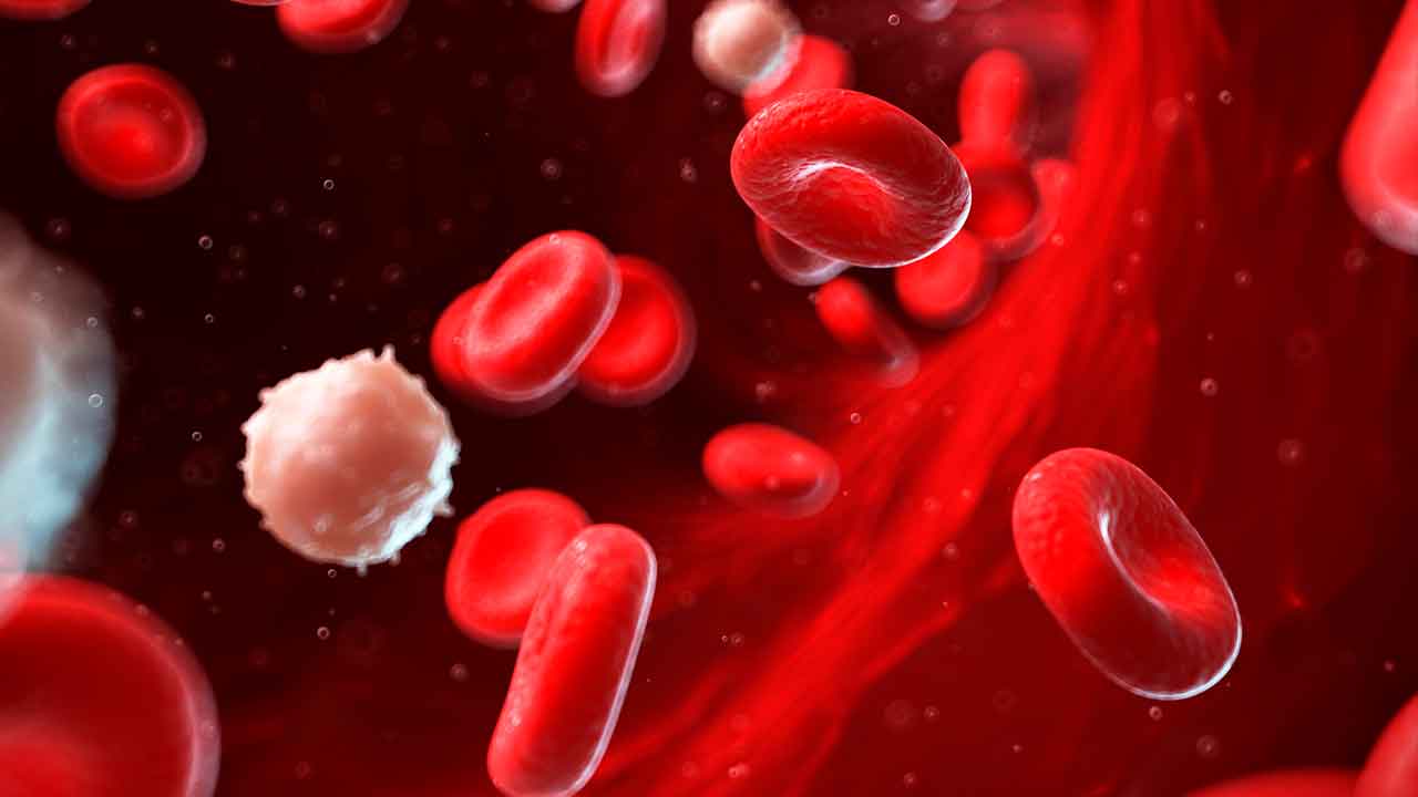
<p class="spai-bg-prepared">All of the cells in our blood – including red cells, white cells and platelets – develop in a process called haematopoiesis from haematopoietic stem cells in our bone marrow. These stem cells are what’s known as multipotent progenitor cells, which simply means that they can develop into more than one cell type.</p> <p class="spai-bg-prepared">Researchers were interested in better understanding how this process changes as we age, so they sequenced the entire genomes of 3,579 haematopoietic stem cells from a total of 10 people – ranging in age from newborn to 81 years.</p> <div class="newsletter-box spai-bg-prepared"> <div id="wpcf7-f6-p193434-o1" class="wpcf7 spai-bg-prepared" dir="ltr" lang="en-US" role="form"> </div> </div> <p class="spai-bg-prepared">Using this information, they were able to construct something similar to a family tree (<a class="spai-bg-prepared" href="https://www.nature.com/scitable/topicpage/reading-a-phylogenetic-tree-the-meaning-of-41956/#:~:text=A%20phylogenetic%20tree%2C%20also%20known,genes%20from%20a%20common%20ancestor." target="_blank" rel="noreferrer noopener">a phylogenetic tree</a>) for each stem cell, showing how the relationships between blood cells changes over the human lifespan.</p> <p class="spai-bg-prepared">They found that in adults under 65, blood cells were produced from between 20,000 and 200,000 different stem cells – each contributing roughly equal amounts to production.</p> <p class="spai-bg-prepared">But after 70 years of age they observed a dramatic decrease in the diversity of stem cells responsible for haematopoiesis in the bone marrow. In fact, only 12-18 independent expanded sets of stem cell clones accounted for 30-60% of cell production.</p> <p class="spai-bg-prepared">These highly active stem cells had outcompeted others and progressively expanded in numbers (clones) across that person’s life, and this expansion (called <a class="spai-bg-prepared" href="https://www.nature.com/articles/s41586-022-04785-z" target="_blank" rel="noreferrer noopener">clonal haematopoiesis</a>) was caused by a rare subset of mutations known as driver mutations that had occurred decades earlier.</p> <p class="spai-bg-prepared">“Our findings show that the diversity of blood stem cells is lost in older age due to positive selection of faster-growing clones with driver mutations. These clones ‘outcompete’ the slower growing ones,” explains lead researcher Dr Emily Mitchell, a haematology registrar at Addenbrooke’s Hospital,UK, and PhD student at the Wellcome Sanger Institute, US.</p> <p class="spai-bg-prepared">“In many cases this increased fitness at the stem cell level likely comes at a cost – their ability to produce functional mature blood cells is impaired, so explaining the observed age-related loss of function in the blood system.”</p> <p class="spai-bg-prepared">Which clones became the dominant stem cells varied between individuals, which explains why variation is seen in disease risk and other characteristics in older adults.</p> <p class="spai-bg-prepared">“Factors such as chronic inflammation, smoking, infection and chemotherapy cause earlier growth of clones with cancer-driving mutations. We predict that these factors also bring forward the decline in blood stem cell diversity associated with ageing,” says joint-senior author Dr Elisa Laurenti, assistant professor at the Wellcome-MRC Cambridge Stem Cell Institute, UK.</p> <p class="spai-bg-prepared">“It is possible that there are factors that might slow this process down, too,” she adds. “We now have the exciting task of figuring out how these newly discovered mutations affect blood function in the elderly, so we can learn how to minimise disease risk and promote healthy ageing.”</p> <p><img id="cosmos-post-tracker" class="spai-bg-prepared" style="opacity: 0; height: 1px!important; width: 1px!important; border: 0!important; position: absolute!important; z-index: -1!important;" src="https://syndication.cosmosmagazine.com/?id=193434&amp;title=Genetic+mutations+slowly+accumulated+over+a+lifetime+change+blood+production+after+70+years+of+age" width="1" height="1" /></p> <div id="contributors"> <p><em><a href="https://cosmosmagazine.com/science/mutations-change-blood-production/" target="_blank" rel="noopener">This article</a> was originally published on <a href="https://cosmosmagazine.com" target="_blank" rel="noopener">Cosmos Magazine</a> and was written by <a href="https://cosmosmagazine.com/contributor/imma-perfetto" target="_blank" rel="noopener">Imma Perfetto</a>. Imma Perfetto is a science writer at Cosmos. She has a Bachelor of Science with Honours in Science Communication from the University of Adelaide.</em></p> <p><em>Image: Getty Images</em></p> </div>

Body

Placeholder Content Image

“Benjamin Button” mice could pave way for reverse ageing

<p>If the three blind mice from the iconic nursery rhyme were living in molecular biologist Dr David Sinclair’s lab at Harvard Medical School, they might not be blind for very long.</p> <p>Dr Sinclair and his team at Harvard Medical School have been using proteins that can turn adult cells into stem cells - a kind of cell that can be turned into any of the specialised cells our bodies need.</p> <p>These stem cells have been helping restore the sight of old mice with damaged retinas, essentially making them younger versions of themselves.</p> <p>“It’s a permanent reset, as far as we can tell, and we think it may be a universal process that could be applied across the body to reset our age,” Dr Sinclair said about his research, which was published in late 2020.</p> <p>The Australian scientist has spent the past 20 years studying ways to reverse the effects of ageing - including the diseases that can afflict us as we get older.</p> <p>“If we reverse ageing, these diseases should not happen,” he said.</p> <p>During a health and wellness talk at Life Itself, Dr Sinclair said the technology is available and it’s only a matter of when we decide to use it.</p> <p>“We have the technology today to be able to go into your hundreds without worrying about getting cancer in your 70s, heart disease in your 80s and Alzheimer’s in your 90s,” he said.</p> <p>“This is the world that is coming. It’s literally a question of when and for most of us, it’s going to happen in our lifetime.”</p> <p>Whitney Casey, an investor who has partnered with Dr Sinclair to create a DIY biological age test, said the researcher wants to “make ageing a disease”.</p> <p>“His research shows you can change ageing to make lives younger for longer,” she said.</p> <p>Dr Sinclair said that when it comes to how modern medicine addresses sickness, it doesn’t tackle the underlying cause, which is usually “ageing itself”.</p> <p>“We know that when we reverse the age of an organ like the brain in a mouse, the diseases of ageing then go away. Memory comes back, there is no more dementia,” he continued.</p> <p>“I believe that in the future, delaying and reversing ageing will be the best way to treat the diseases that plague most of us.”</p> <p>Dr Sinclair’s research comes amid a global effort by scientists working to reprogram adult cells into stem cells, started by Japanese researcher Shinya Yamanaka, who won a Nobel Prize for reprogramming adult skin cells into behaving like embryonic (or pluripotent) stem cells.</p> <p>These “induced pluripotent stem cells” became known as “Yamanaka factors”, with later research finding that exposing cells to four of the main Yamanaka factors could remove signs of ageing.</p> <p>Since their original study, where they discovered that damaged cells were able to be rejuvenated by injecting three of these factors into the eyes of mice, Dr Sinclair and his lab have reversed ageing in mouse brains and muscles, and are now working on a mouse’s whole body.</p> <p>Dr Sinclair said their discovery indicated that there is a “back-up copy” of youthful information stored in the body, which he calls the “information theory of ageing”.</p> <p>“It’s a loss of information that drives ageing cells to forget how to function, to forget what type of cell they are,” he revealed.</p> <p>“And now we can tap into a reset switch that restores the cell’s ability to read the genome correctly again, as if it was young.”</p> <p><em>Image: Getty Images</em></p>

Body

Placeholder Content Image

Woman celebrates 100th birthday in jail cell

<p>Ruth Bryant celebrated her centennial birthday by crossing off a wish on her bucket list: to be arrested and sent to jail.</p> <p>The US woman was celebrating her 100<sup>th</sup> birthday on Wednesday at her assisted living community in North Carolina when deputies from the Person County Sheriff’s Office showed up and served her a warrant for “indecent exposure” at a fire department.</p> <p>Friends and family members present at Bryant’s birthday celebrations weren’t aware of the plan, <em>WRAL </em>reported.</p> <p>“I know that she is a hundred years old, but I didn’t know ... they’d be going this far,” the 100-year-old’s daughter Marian Oakley told the outlet.</p> <p><iframe src="https://www.facebook.com/plugins/video.php?href=https%3A%2F%2Fwww.facebook.com%2FKATVChannel7%2Fvideos%2F2729937517059685%2F&amp;show_text=1&amp;width=560" width="560" height="445" style="border: none; overflow: hidden;" scrolling="no" frameborder="0" allowtransparency="true" allow="encrypted-media" allowfullscreen="true"></iframe></p> <p>Police handcuffed Bryant to her walker and loaded her into the front seat of the police car before driving her to prison.</p> <p>She spent a few minutes inside a cell and was given a free phone call, a mug shot and an orange jail t-shirt.</p> <p>“I’m in the jailhouse now! I finally got here!” she said.</p> <p>She was released after paying bail in the form of a hug to the chief jailer and returned to her residence for cakes with friends.</p>

Retirement Life

Placeholder Content Image

Packing cells will change how you travel in 2019

<p>Packing cells – people either love them or think they are a huge waste of money. We’ve detailed the pros and cons of packing cells so that you can make your own mind up.</p> <p><strong>1. What are they?</strong></p> <p>Packing cells are little cubes or zippered bags of various sizes that act as removable compartments for your suitcase or backpack.</p> <p><strong>2. How do you use them?</strong></p> <p>Packing cells allow you to organise your suitcase. You sort the items you need into individual bags. Put your dirty clothes in one, underpants in another. Put your socks in one, camera gear in another. You get the point. If you’re sharing a suitcase with a travelling companion, you can put your clothes into individual packing cells – that way your clothes won’t get all mixed up.</p> <p><strong>3. What do fans say about them?</strong></p> <p>A Facebook thread on packing cells went viral this week due to the number of people commenting. Comments such as: “Best things ever – saves so much room and keeps things tidy and organised” were common. Here’s a few more comments: “They have really changed our packing. Highly recommend. No more digging through the whole bag trying to find a pair of undies.” – Alicia thoman “We use them all the time now. Each person has their own pack and then you just take it out of the case – so much easier.” – Clare Ditchburn “They are the best, love mine, make so much more room in your suitcase.” – Kathy Stringfellow</p> <p><strong>4. What do the critics say?</strong></p> <p>Critics say that packing bags are a waste of money. Some argue that the bags are just more stuff you don’t need. Why pay the money when it doesn’t really take that long to find something in your bag. Is the 20 seconds really worth the cash?</p> <p><strong>5. Tips for using them</strong></p> <p>Generally, most people we found who have used the packing bags say they love them. So how do you use them effectively?</p> <ul> <li>Use a different colour per traveller</li> <li>Make sure you buy enough of them</li> <li>Get packing bags that have a clear window or mesh to allow you to see what is in the bag. Otherwise you’re going to spend just as much time hunting for the stuff you need.</li> <li>You can make your own packing bags from laundry bags, old airline amenities bags or plastic zip-lock bags.</li> <li>Buy a selection of different sizes</li> <li>Use them for small and necessary items.</li> <li>Use one for medications</li> <li>Keep one for chargers and phones</li> <li>Have a waterproof one for wet clothes</li> <li>Have one for dirty clothes</li> <li>Where do you buy them?</li> </ul> <p><em>Written by Alison Godfrey. Republished with permission of </em><a href="https://www.mydiscoveries.com.au/stories/packing-cells-hack/"><em>MyDiscoveries</em></a><em>.</em></p>

Travel Tips

Placeholder Content Image

Exercise slows ageing of cells

<p>The secret to keeping your body youthful may be found in the way you move.</p> <p>A new study has found that high-intensity interval training (HIIT) can essentially stop cellular ageing in its tracks and, in some cases, rejuvenate the cells that repair damage in the body.</p> <p>For the study, researchers from the Mayo Clinic took 36 men and 36 women split into younger (aged between 18 and 30) and older (aged between 65 and 80) age groups.<br /> <br /> The participants were then assigned a three-month programme of HIIT, strength training or a combination of the two.</p> <p>They already knew that both <a href="http://www.smh.com.au/lifestyle/diet-and-fitness/time-to-get-fit-a-guide-to-hiit-20160518-goxmgm.html">HIIT</a> and <a href="http://www.smh.com.au/lifestyle/health-and-wellbeing/fitness/the-science-of-strength-seven-ways-muscle-makes-us-healthier-20170312-guwcpv.html">strength training</a> provided enormous health benefits to our bodies, they just didn't know exactly how or why, or which was better.</p> <p>So, to understand the way exercise affects us at a molecular level, the researchers then took biopsies from the participants' thigh muscles and compared them with samples from sedentary volunteers.</p> <p>The strength-training group predictably saw the greatest improvements in muscle mass, but the findings that have been described as "<a href="https://www.psychologytoday.com/blog/the-athletes-way/201703/mayo-clinic-study-identifies-how-exercise-staves-old-age">earth shattering</a>" were at a cellular level in the HIIT group. </p> <p>Mitochondria are the "<a href="https://www.umdf.org/what-is-mitochondrial-disease/">powerhouses" of our cells</a>, responsible for creating more than 90 per cent of the energy needed by the body to sustain life and support organ function. Their function typically declines with age.  </p> <p>However, in the HIIT group, the mitochondrial functioning improved by 69 per cent among the older participants, and by 49 per cent among the younger group. </p> <p>As well as improving their insulin levels, heart and lung health, some in the high-intensity biking group also saw a reversal of the age-related decline in mitochondrial function and proteins needed for building muscle.</p> <p>The research provided an explanation for the many health benefits of exercise said the lead senior author, Sreekumaran Nair.</p> <p>"Based on everything we know, there's no substitute for these exercise programmes when it comes to delaying the ageing process," says Nair, of the study published in the journal <a href="http://www.cell.com/cell-metabolism/abstract/S1550-4131(17)30099-2?_returnURL=http%3A%2F%2Flinkinghub.elsevier.com%2Fretrieve%2Fpii%2FS1550413117300992%3Fshowall%3Dtrue">Cell</a>. "These things we are seeing cannot be done by any medicine."</p> <p>He adds: "If people have to pick one exercise, I would recommend high-intensity interval training, but I think it would be more beneficial if they could do three to four days of interval training and then a couple days of strength training."  </p> <p>Emmanuel Stamatakis of the Charles Perkins Centre at the University of Sydney says it is a "fascinating piece of research".</p> <p>"This not only sheds light on how high-intensity interval exercise works at the cellular level, but [also] on the potential of vigorous exertion in general," says Stamatakis, who was not involved with the research. </p> <p>It also shows what is happening beneath the sweat that makes HIIT more beneficial to our bodies than other forms of exercise. Which aspect of HIIT is responsible for such dramatic changes, however, is still an unknown.</p> <p>"Assuming that the key attribute of HIIT is the vigorous intensity that challenges the human physiology to make rapid adaptations, this research supports well what we saw recently <a href="http://jamanetwork.com/journals/jamainternalmedicine/article-abstract/2596007">in a large epidemiologic study</a> where even one to two sessions per week of  predominantly sport/ exercise of vigorous intensity were associated with substantial all-cause, CVD and cancer mortality benefits," Stamatakis explains.</p> <p>"These benefits were comparable with meeting the physical activity recommendations by doing regular physical activity of mostly moderate intensity."</p> <p>Now the question is whether HIIT is right for everyone. Given how few of us manage to meet the recommendations, Stamatakis remains unsure.</p> <p>"There <a href="https://ijbnpa.biomedcentral.com/articles/10.1186/s12966-015-0254-9">is a big debate</a> as to whether HIIT is the way to go for better population health, but it is certain that it has a time and a place," he says. "Although not every physically inactive person would be willing or able to join a HITT program, this new piece of research highlights that in addition to  public health messages like 'move as often as possible, a little is better than nothing', we need to also add 'aim to huff and puff sometimes'."</p> <p>This might be as simple as taking the stairs whenever you can – both a form of incidental and HIIT exercise. </p> <p>"For many people, stair climbing will involve bouts of high-intensity activity lasting one or more minutes, and if this is repeated regularly enough in everyday life it could potentially improve fitness and other aspects of cardiovascular and metabolic health quite rapidly," Stamatakis says.   </p> <p>And of course, it will keep us young.</p> <p>"There are substantial basic science data to support the idea that exercise is critically important to prevent or delay ageing," says Nair, who plans to look at the effect of exercise on other tissues in the body. "There's no substitute for that."</p> <p><em>Written by Sarah Berry. Appeared on <span style="text-decoration: underline;"><strong><a href="http://www.stuff.co.nz/" target="_blank">Stuff.co.nz.</a><br /></strong></span></em></p>

Body

Placeholder Content Image

Crackdown on drivers using cell phones

<p>A record number of drivers have been caught paying more attention to their cellphones than to the road.</p> <p>Police say a West Aucklander with a beer in one hand and a phone in the other was among the 4195 drivers caught and fined a total of $331,680 during a crackdown in September.</p> <p>The latest police figures show a more than threefold increase in the number of drivers caught nationwide on their phones that month, compared with the average monthly numbers.</p> <p>In the entire Auckland region, 1932 drivers were caught on their phones in September, compared with 963 in August. </p> <p>The number of Wellingtonians snapped increased 75 per cent to 273, and the number of Cantabrians caught almost doubled, with 830 drivers fined.</p> <p>Road policing national manager Superintendent Steve Greally said the figures were horrifying. "People are just flouting it. They don't understand the risk of taking your eyes off the road even for just two seconds.</p> <p>"Even for just a second, you could kill a cyclist, another driver – even yourself. It's a huge issue for us."</p> <p>Greally said he understood how drivers idling in gridlock or at a red light might think it was safe to use their phones, but it was illegal because it was risky.</p> <p>"Even if you're going 5 or 6 kilometres per hour, you're going to cause a nose-to-tail. Or a small child might run out in front of you who doesn't know any better."</p> <p>A record number of drivers were fined in September for using mobile phones while driving.</p> <p>He said officers had not been given quotas during the two-week September sting, but all officers, including detectives who did not usually do road policing, were asked to pull over drivers on cellphones when they spotted them.</p> <p>Breaking the rules results in an $80 fine and 20 demerit points. If 100 points are accumulated within two years, drivers automatically lose their licences for three months.</p> <p>Greally said he was unaware of anyone who had lost their licence purely for repeated phone offences, but thought it likely.</p> <p>"With technology, you've getting a lot more things that you can do with your phone.</p> <p>"They are not just used for calling or for texting. It's apps – the more that increases, the more we'll be having people using phones while driving."</p> <p>The Automobile Association congratulated police on the crackdown, saying multi-tasking impaired drivers' judgment.</p> <p>"There's no doubt it takes your attention away from your driving and increases your risk," spokesman Dylan Thomsen said.</p> <p>"There's a reasonable difference between talking on your phone and to passengers who are in the same environment as the driver and more likely to realise what is going on. Passengers tend to mute their conversation." </p> <p>As many people's phones doubled as music players, the AA's interpretation was that "infrequently" touching a phone attached to a car stereo to change a song would not be breaking the law, any more than changing the radio station would be, Thomsen said.</p> <p>Ministry of Transport figures from 2014 showed distracted drivers were a factor in 12 per cent of crashes.</p> <p>Distracted driving killed 22 people, seriously injured 191, and chalked up an estimated $297 million in social costs.</p> <p>Between 2011-13, 163 fatal or injury crashes were attributed to drivers using cellphones.</p> <p>Backing up the NZ data, a new report out of the US has suggested that smartphones and other driving distractions could be making roads there more dangerous.</p> <p>Preliminary stats released Thursday (NZ time) showed US road deaths for January-June 2015 had risen 8.1 per cent to 16,225 which is a rate more than double an increase  in overall driving spawned by falling fuel prices and a growing economy.</p> <p>"The increase in smartphones in our hands is so significant, there's no question that has to play some role. But we don't have enough information yet to determine how big a role," said Mark Rosekind, who heads the National Highway Traffic Safety Administration, the US government's auto safety watchdog.</p> <p>While US officials said it was too early to identify contributing factors. But Rosekind told reporters that officials are looking at likely causes including distracted driving and the possibility lower fuel prices have encouraged more driving among "risky drivers" such as teenagers.</p> <p>Unlike NZ's laws, Rosekind also criticised an absence of effective laws in US states that prohibit hand-held smartphones by drivers or require the use of seatbelts and motorcycle helmets.</p> <p>The New Zealand Transport Agency has even looked at independent research into hi-tech mobile phone detectors as it considered whether to nationally trial the devices in 2014.</p> <p>They are designed to detect transmissions from passing drivers' phones and sort them from the background noise of other phones in the area.</p> <p>The researchers concluded the detectors would be useful alongside visual observation – but there were concerns the detectors might pick up emails, calls and texts being received by a phone without the driver touching it.</p> <p>Written by Talia Shadwell. First appeared on <a href="http://www.Stuff.co.nz" target="_blank"><span style="text-decoration: underline;"><strong>Stuff.co.nz</strong></span></a>.</p>

News

Placeholder Content Image

Stems cells could be used to treat macular degeneration

<p>A study at Cedars-Sinai Medical Center in Los Angeles has found that an injection of stem cells in the eyes could be an effective treatment for vision loss caused by age-related macular degeneration. Currently, there is no treatment that slows the progression of the disease, which is the leading cause of vision loss in people over 65.</p> <p>The study’s lead author, Shaomei Wang, MD, PhD explained, “This is the first study to show preservation of vision after a single injection of adult-derived human cells into a rat model with age-related macular degeneration.” Published in the journal STEM CELLS, the study resulted in 130 days of preserved vision in laboratory rat; this equates roughly to 16 years for a human.</p> <p>When animals with macular degeneration were injected with stem cells created using adult skin cells, healthy cells began to migrate around the retina, forming a protective layer. This shield prevented ongoing degeneration of the vital retinal cells responsible for vision.</p> <p>For those who don’t know, age-related macular degeneration occurs when the small central portion of the retina – the macula – deteriorates. Aside from age, other causes of macular degeneration include a genetic predisposition, and environmental factors.</p> <p>The next steps in the process include testing the safety of the stem cell injection in preclinical animal studies. In the future, clinical trials will be designed to test any potential benefit in patients with later-stage age-related macular degeneration.</p> <p><span style="text-decoration: underline;"><strong><a href="http://www.sciencedaily.com/releases/2015/04/150414093554.htm" target="_blank">Source: Science Daily</a></strong></span></p>

Eye Care