Placeholder Content Image

Son's first dance with mum suffering from motor neurone disease

<p>An emotional wedding video of a wheelchair-bound mother severely affected by motor neurone disease (MND) dancing with her eldest son on his big day has gone viral, with people describing the moment as both heart-breaking and heart-warming at the same time. </p> <p>Kathy Poirer was watching her eldest son Zak get married in a moving ceremony in their native Florida. </p> <p>Despite being unable to walk, Kathy, with the help of her other two children, was able to dance with her eldest child on his big day. </p> <p>The moment was captured on video and has since gone viral, as Kathy said the dance was a dream come true. </p> <p>“I just really wanted that moment with my son,” Poirer told <em>Sunrise</em>. </p> <p>“There are so many milestones in their life and you look forward to them and I was afraid I wouldn’t have that opportunity."</p> <p>“It truly is an expression of a mother and her son and their love for each other.”</p> <p>Kathy was diagnosed with MND more than three years ago, and has bravely fought the diagnosis and challenged the odds against her ever since.</p> <p>“I prayed every day that I would make it long enough to dance with him, and I fight every day for my life,” she said.</p> <p>Motor neurone disease is a condition that affects the body’s brain and the nerve cells called motor neurones.</p> <p>According to <a href="https://www.mndaustralia.org.au/mnd-connect/what-is-mnd/what-is-motor-neurone-disease-mnd" target="_blank" rel="noopener" data-link-type="article-inline">MND Australia:</a> “Motor neurones normally carry messages from the brain to the muscles via the spinal cord. The messages allow people to make voluntary movements like walking, swallowing, talking and breathing."</p> <p>There is currently no cure for MND, and the condition is known to drastically reduce life expectancy. </p> <p>Kathy is devoting the rest of her life to bringing awareness to this devastating disease, while encouraging people not to take life for granted. </p> <p>“What I wanted to do was make people aware that this can happen to you in a heartbeat and just live every moment to the fullest - you never know when it’s gonna get taken away,” she said.</p> <p><em>Image credits: Sunrise</em></p>

Caring

Placeholder Content Image

Oops! Scientists identify the neurons responsible for learning from mistakes

<p>Have you ever driven past an intersection and registered you should have turned right a street ago, or been in a conversation and, as soon as the words are out of your mouth, realised you really shouldn’t have said that thing you just did?</p> 
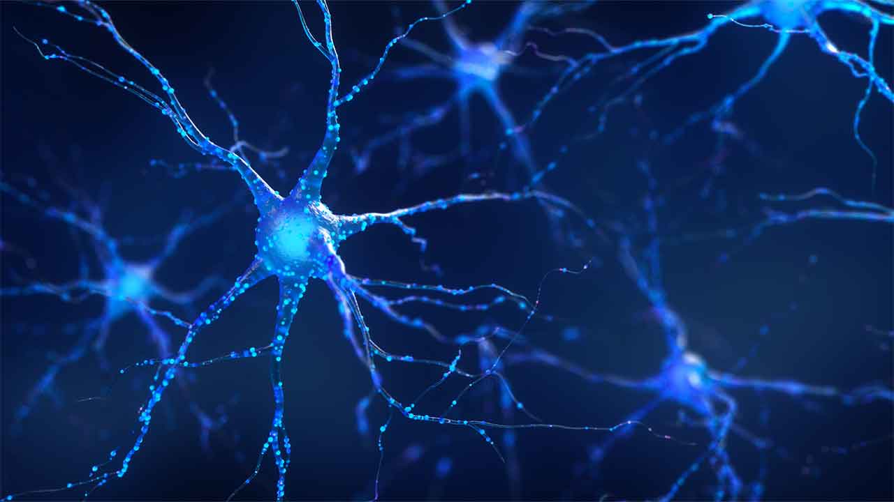
<p>It’s a phenomenon known as performance monitoring; an internal signal produced by the brain that lets you know when you’ve made a mistake.</p> <p>Performance monitoring is a kind of self-generated feedback that’s essential to managing our daily lives. Now, neuroscientists have discovered that signals from <a href="https://cosmosmagazine.com/science/biology/brain-pleasers-the-neurons-that-respond-to-singing/" target="_blank" rel="noreferrer noopener">neurons</a> in the brain’s medial frontal cortex are responsible for it.</p> <p>A <a href="https://www.science.org/doi/10.1126/science.abm9922" target="_blank" rel="noreferrer noopener">new study</a> published in <em>Science</em> reports that these signals are used to give humans the flexibility to learn new tasks and the focus to develop highly specific skills.</p> <p>“Part of the magic of the human brain is that it is so flexible,” says senior author Ueli Rutishauser, professor of Neurosurgery, Neurology, and Biomedical Sciences at Cedars-Sinai Medical Center, US. “We designed our study to decipher how the brain can generalise and specialise at the same time, both of which are critical for helping us pursue a goal.”</p> <p>They found that the performance monitoring signals help improve future attempts of a particular task by passing information to other areas of the brain. They also help the brain adjust its focus by signalling how much conflict or difficulty was encountered during the task.</p> <p>“An ‘Oops!’ moment might prompt someone to pay closer attention the next time they chat with a friend, or plan to stop at the store on the way home from work,” explains first author Zhongzheng Fu, researcher in the Rutishauser Laboratory at Cedars-Sinai.</p> <p>The team recorded the activity of more than 1000 neurons in the medial frontal cortexes of human epilepsy patients (who had existing electrode brain implants to help locate the focus of their seizures) while they performed complex cognitive tasks.</p> <div class="newsletter-box"> <div id="wpcf7-f6-p190553-o1" class="wpcf7" dir="ltr" lang="en-US" role="form"> </div> </div> <p>In the first task, called the Stroop task, participants’ reading- and colour naming skills were tested. Viewing the written name of the colour, such as “red”, printed in the ink of a different colour, such as blue, they were asked to name the ink colour rather than the written word.</p> <p>In the second task – the Multi-Source Interference Task (MSIT) – participants were shown three digits on a screen (two the same number and the other unique) and had to press a button associated with the unique number while resisting the tendency to press the other (because it appears twice).</p> <p>The researchers noted that two types of neurons seemed to be at work: “error” neurons fired strongly after a mistake was made, while “conflict” neurons fired in response to the difficulty of the task.</p> <p>“When we observed the activity of neurons in this brain area, it surprised us that most of them only become active after a decision or an action was completed,” says Fu. “This indicates that this brain area plays a role in evaluating decisions after the fact, rather than making them.”</p> <p>Scientists have known for some time that there are two types of performance monitoring: domain general and domain specific.</p> <p>Domain general performance monitoring tells us <em>when</em> something goes wrong, which allows people to perform new tasks with little instruction. Domain specific monitoring tells them <em>what</em> went wrong, and is one way that people perfect individual skills.</p> <p>Previously it was thought that the different neurons responsible for these two forms were located in distinct parts of the brain, but this research has found that they’re actually intermingled in the medial frontal cortex.</p> <p>According to Rutishauser, understanding the mechanisms behind performance monitoring is critical to perfecting the treatment of certain psychiatric disorders in which it is extreme, for example obsessive compulsive disorder (overactive monitoring) and schizophrenia (underactive).</p> <p><img id="cosmos-post-tracker" style="opacity: 0; height: 1px!important; width: 1px!important; border: 0!important; position: absolute!important; z-index: -1!important;" src="https://syndication.cosmosmagazine.com/?id=190553&title=Oops%21+Scientists+identify+the+neurons+responsible+for+learning+from+mistakes." width="1" height="1" data-spai-target="src" data-spai-orig="" data-spai-exclude="nocdn" /></p> <div id="contributors"> <p><em><a href="https://cosmosmagazine.com/science/biology/neurons-performance-monitoring/" target="_blank" rel="noopener">This article</a> was originally published on <a href="https://cosmosmagazine.com" target="_blank" rel="noopener">Cosmos Magazine</a> and was written by <a href="https://cosmosmagazine.com/contributor/imma-perfetto" target="_blank" rel="noopener">Imma Perfetto</a>. Imma Perfetto is a science writer at Cosmos. She has a Bachelor of Science with Honours in Science Communication from the University of Adelaide.</em></p> <p><em>Image: Getty Images</em></p> </div>

Body

Placeholder Content Image

How efficient is the human brain?

<p><span style="font-size: 14px;">What is the difference between human brains and those of other mammals? Potentially, energy efficiency, according to a </span><a style="font-size: 14px;" rel="noopener" href="https://dx.doi.org/10.1038/s41586-021-04072-3" target="_blank">study</a><span style="font-size: 14px;">, published in </span><em style="font-size: 14px;">Nature.</em></p> <div class="copy"> <p>Neurons in the brain communicate with each other via electrical pulses. These pulses, generated as ions such as potassium and sodium, enter the cell through an ion channel. The channels act as an “on-off” switch by changing shape to alter the flow of the ions, either letting them in and generating an electrical signal, or closing to prevent the signal getting through.</p> <p>Now, a team of researchers from Massachusetts Institute of Technology have found that the number of ion channels in the human brain are at a lower density than in other mammals’ brains. They have hypothesised that this has helped human brains become more efficient.</p> <p>The researchers analysed neurons from 10 mammals – the most extensive electrophysical study of its kind to date – and found that bigger neurons lead to more ion channels in a relatively constant ratio of size-to-channels.</p> <p>What this means is that bigger neurons can still handle all the electrical pulses they are bombarded with because they have the ion channel capacity to process the ions. No matter the brain size, the energetic cost to run each mammal’s brain was about the same.</p> <p><strong>The human brain is the exception</strong></p> <p>That is, in every mammal except humans, who had a much lower density of ion channels than expected.</p> <p>“Previous comparative studies established that the human brain is built like other mammalian brains, so we were surprised to find strong evidence that human neurons are special,” says former MIT graduate student Lou Beaulieu-Laroche.</p> <p>Graphical abstract. Created on imgflip by Cosmos.</p> <p>One benefit of a lower density of ion channels may be that less energy is used on pumping ions into neurons, which could then be diverted to other processes, like creating more complicated synaptic connections.</p> <p>“If the brain can save energy by reducing the density of ion channels, it can spend that energy on other neuronal or circuit processes,” says Mark Harnett, an associate professor of brain and cognitive sciences, a member of MIT’s McGovern Institute for Brain Research, and the senior author of the study.</p> <p>“We think that humans have evolved out of this building plan that was previously restricting the size of cortex, and they figured out a way to become more energetically efficient, so you spend less [energy] per volume compared to other species.”</p> <p>In this case, the human brain could run on the same amount of power as other mammal brains but perform more complex procedures with the excess energy diverted from ion channels.</p> <p>Harnett hopes to study where that extra energy is being used and whether there are any specific genes responsible for ion channel density exclusive to humans.</p> <!-- Start of tracking content syndication. Please do not remove this section as it allows us to keep track of republished articles --> <img id="cosmos-post-tracker" style="opacity: 0; height: 1px!important; width: 1px!important; border: 0!important; position: absolute!important; z-index: -1!important;" src="https://syndication.cosmosmagazine.com/?id=172198&amp;title=How+efficient+is+the+human+brain%3F" alt="" width="1" height="1" /> <!-- End of tracking content syndication --></div> <div id="contributors"> <p><em><a rel="noopener" href="https://cosmosmagazine.com/science/biology/how-efficient-is-the-human-brain/" target="_blank">This article</a> was originally published on <a rel="noopener" href="https://cosmosmagazine.com" target="_blank">Cosmos Magazine</a> and was written by <a rel="noopener" href="https://cosmosmagazine.com/contributor/dr-deborah-devis" target="_blank">Deborah Devis</a>. Deborah Devis is a science journalist at Cosmos. She has a Bachelor of Liberal Arts and Science (Honours) in biology and philosophy from the University of Sydney, and a PhD in plant molecular genetics from the University of Adelaide.</em></p> <p><em>Image: Getty Images</em></p> </div>

Mind

Placeholder Content Image

New study offers hope to those with motor neurone disease

<p><span style="font-weight: 400;">Motor neurone disease, also called amyotrophic lateral sclerosis (ALS), is a rare condition that slowly kills off nerve cells in the brain and spine, leading to paralysis and eventually death.</span></p> <p><span style="font-weight: 400;">Though currently incurable, </span><a rel="noopener" href="https://academic.oup.com/braincomms/article/3/3/fcab166/6340444" target="_blank"><span style="font-weight: 400;">a new study</span></a><span style="font-weight: 400;"> may have brought us closer to finding one.</span></p> <p><span style="font-weight: 400;">Scientists have taken lab samples of one form of ALS and been able to reverse one of the hallmark biological abnormalities the disease introduces in cells.</span></p> <p><span style="font-weight: 400;">Although this is yet to be applied in other forms of the disease or in human beings, the finding represents a large step forward in understanding how ALS could be combatted, providing some hope that the disease could one day be beaten.</span></p> <p><strong>What the study found</strong></p> <p><span style="font-weight: 400;">The researchers found that three RNA binding proteins, which help regulate RNA, get stuck in the wrong place for most people with ALS.</span></p> <p><span style="font-weight: 400;">Instead of staying in the motor neuron nucleus, they end up in the surrounding cytoplasm.</span></p> <p><span style="font-weight: 400;">The team then found that blocking a particular enzyme, called VCP, was able to reverse this in their human cell samples and return the distribution of RNA binding proteins in the nucleus and cytoplasm back to normal.</span></p> <p><span style="font-weight: 400;">According to the scientists, this suggests that this enzyme becomes mutated and overactive in some cases of ALS.</span></p> <p><span style="font-weight: 400;">“Demonstrating proof-of-concept for how a chemical can reverse one of the key hallmarks of ALS is incredibly exciting,” said Jasmine Harley, a neuroscientist from the Francis Crick Institute in the UK.</span></p> <p><span style="font-weight: 400;">“We showed this worked on three key RNA binding proteins, which is important as it suggests it could work on other disease phenotypes too.”</span></p> <p><span style="font-weight: 400;">The drug they used to inhibit the enzyme is also being tested in cancer trials, which could speed up its development and availability if it is found to help cancer patients and ALS patients.</span></p> <p><span style="font-weight: 400;">In a second study, published in the journal </span><em><a rel="noopener" href="https://academic.oup.com/brain/advance-article/doi/10.1093/brain/awab078/6164957" target="_blank"><span style="font-weight: 400;">Brain</span></a></em><span style="font-weight: 400;">, the same researchers found over 100 types of RNA fragments, called intron-retaining transcripts, which can also move from the nucleus of cells into the cytoplasm in ALS cases.</span></p> <p><span style="font-weight: 400;">The researchers found that these fragments have sequences that bind to the RNA binding proteins, and they suspect these sequences are drawing the proteins out of the nucleus and into the cytoplasm.</span></p> <p><img style="width: 0px; height: 0px;" src="/nothing.jpg" alt="" data-udi="umb://media/1f84fdb6755a48e884e50245585f3db4" /><img style="width: 500px; height: 433.3333333333333px;" src="https://oversixtydev.blob.core.windows.net/media/7843072/gettyimages-1129371731.jpg" alt="" data-udi="umb://media/1f84fdb6755a48e884e50245585f3db4" /></p> <p><em><span style="font-weight: 400;">Image: Getty Images</span></em></p> <p><span style="font-weight: 400;">“To imagine what’s going on here we can consider watching a movie at the cinema,” neuroscientist Jacob Neeves explained.</span></p> <p><span style="font-weight: 400;">“Typically, we don’t expect to see adverts throughout the film, but, if something goes wrong these ads might start cropping up at odd and unexpected points. These retained introns are a little bit like these abnormal ad breaks.”</span></p> <p><strong>Why this matters</strong></p> <p><span style="font-weight: 400;">Though only 1-2 percent of ALS cases have the mutated enzyme the scientists found in the first study, both pieces of research add to our understanding of motor neurone disease.</span></p> <p><span style="font-weight: 400;">This additional information offers new hope that scientists could eventually figure out how to undo some of the damage that the disease causes to the brain and nervous system.</span></p> <p><span style="font-weight: 400;">“More research is needed to investigate this further,” Harley said. “We need to see if this might reverse other pathological hallmarks of ALS and also, in other ALS disease models.”</span></p>

Mind